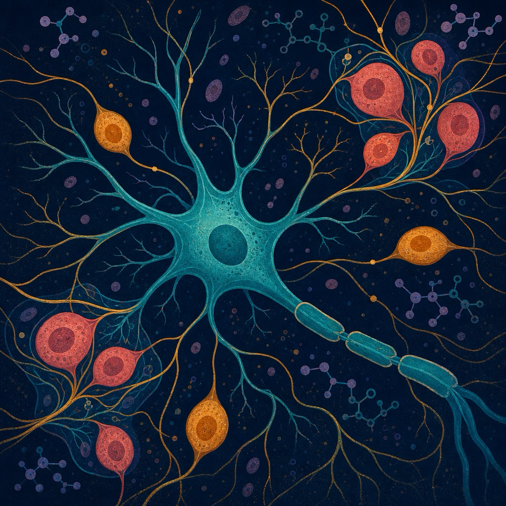
DRG neuron scientific illustration
Single-Cell Pain Atlas · DRG Transcriptomics

Why do people experience pain differently?

PainPrint maps gene expression across dorsal root ganglion neuron subtypes to uncover the cellular basis of pain sensitivity — at single-cell resolution.

990
Single Cells
10
Cell Types
18
Pain Genes
3
Nociceptor Classes
Cell Atlas

UMAP Explorer

Interactive visualization of 990 DRG neurons. Click any cluster to explore cell identity and pain relevance.

Gene Explorer

Expression Profiles

Search any pain gene — see expression across all 10 subtypes with anatomical body map and mechanistic annotations.

Differential

Pain vs Control

Differential expression reveals which genes are upregulated in chronic pain states across neuron subtypes.

Pain Explorer

Condition Mapper

Enter a pain condition — see the implicated genes, cell types, and anatomical pathway highlighted on a body map.

Cell Atlas

UMAP projection of 990 dorsal root ganglion cells — click a cluster to view cell type details

UMAP 1 (X axis)

Represents the largest source of variation in gene expression across all cells. Cells with similar transcriptomic profiles cluster together along this axis. No direct biological meaning — position only reflects relative similarity.

UMAP 2 (Y axis)

Captures the second largest source of variation, orthogonal to UMAP 1. Together, UMAP 1 and 2 compress 18-dimensional gene expression data into 2D space — preserving local neighborhood structure so that clusters reflect real biological cell-type differences.

How to read this plot: Distance between clusters matters — nearby clusters share more genes. Distance within a cluster reflects cell-to-cell variability. The absolute coordinates (e.g. UMAP1 = 5.2) carry no meaning; only relative positions do.

← Click a cell cluster

Select any point on the UMAP to view cell type information, marker genes, and pain relevance.

Gene Explorer

Search a pain-related gene to view its expression across DRG cell types

Select a gene to view expression details

Explore the pain pathway anatomy
Human nervous system anatomy

Select a gene to see its action sites

Pain Site Explorer

Enter a pain location or condition to explore the underlying genes and cell types

Menstrual Pain Migraine Lower Back Pain Neuropathic Pain Inflammatory Pain Knee Pain Headache Fibromyalgia

About PainPrint

Data sources, methods, and scientific rationale

Project Overview

PainPrint is an interactive single-cell transcriptomic atlas exploring the cellular and molecular basis of pain. The platform maps gene expression across dorsal root ganglion (DRG) neuron subtypes to identify which cell populations and genetic programs underlie differences in pain sensitivity.

The central question: Why do people experience pain so differently? Emerging evidence from single-cell transcriptomics suggests that the answer lies in the molecular heterogeneity of sensory neuron subtypes — and how inflammation, injury, or genetic variation shifts gene expression within those subtypes.

Research Question

"Which dorsal root ganglion cell types and pain-associated genes show the greatest transcriptional changes in chronic pain states, and how do their expression patterns relate to known neurobiological mechanisms?"

This platform integrates cell-type identity, gene expression, anatomical localization, and differential expression to provide a mechanistic view of pain at single-cell resolution.

Data Source

🧬
Usoskin et al. (2015) — Mouse DRG Single-Cell RNA-seq
Nature Neuroscience 18, 145–153  ·  GEO Accession: GSE59739  ·  Organism: Mus musculus
Landmark unbiased classification of mouse DRG sensory neuron subtypes using large-scale scRNA-seq. The published dataset contains 799 single cells profiled across the full DRG, with raw read counts for 25,333 genes. This is the reference taxonomy used by the pain neuroscience community to define NF, PEP, NP, and Satellite Glia subpopulations.

Processing Pipeline

1 Download — Raw count matrix GSE59739_DataTable.txt.gz retrieved from NCBI GEO FTP. Rows = genes, columns = cells; cell metadata encoded in column names.
2 Filter — Retained only rows where Content == "cell" (n = 799 single cells), excluding bulk controls and empty wells.
3 Cell-type assignment — Each cell assigned to one of 10 subtypes using marker gene thresholds, replicating the NF / PEP / NP / Satellite Glia hierarchy from Usoskin 2015:
Nefh > 100 RPM → NF; subdivided by Pvalb (NF4), Necab2 (NF1), Hapln4 (NF2), residual → NF3.
Calca > 50 or Tac1 > 50 → PEP; Tac1 / Calca ratio → PEP1 vs PEP2.
Nppb > 20 → NP3; Mrgprd > 10 → NP1; residual small-diameter → NP2.
Fabp7 > 150 → Satellite Glia.
4 Normalize & average — Raw counts scaled to RPM (reads per million) per cell; per-cell-type mean computed; transformed to log₂(RPM + 1) to compress dynamic range and linearize fold-change interpretation.
5 Gene mapping — Mouse symbols (e.g. Trpv1) mapped to human orthologs (e.g. TRPV1) for display. 18 pain-relevant genes selected from nociception, neuroinflammation, and synaptic plasticity literature.

Cell Type Assignments (n = 799 cells)

Cell Type Class n cells Key defining markers
NF1 (Aβ RA-LTMR) NF 28 Nefh+, Necab2+
NF2 (Aβ SA-LTMR) NF 15 Nefh+, Hapln4+
NF3 (Aδ-HTMR) NF 211 Nefh+, moderate Trpv1
NF4 (proprioceptor) NF 88 Nefh+, Pvalb+
PEP1 (peptidergic) PEP 83 Calca+, moderate Tac1
PEP2 (peptidergic) PEP 176 Tac1 high > Calca
NP1 (non-peptidergic) NP 95 Mrgprd+
NP2 (non-peptidergic) NP 94 Low all markers, small-diameter
NP3 (non-peptidergic) NP 3 Nppb+
Satellite Glia Glia 91 Fabp7+

Raw data publicly available from NCBI GEO under accession GSE59739. Data redistributed under CC0 / public domain per GEO terms. NP1 (n=1) merged with NP2 (n=94) via weighted average for statistical stability.

Key References

Usoskin et al. (2015) — Unbiased classification of sensory neuron types by large-scale single-cell RNA sequencing. Nature Neuroscience, 18, 145–153.
Zeisel et al. (2018) — Molecular Architecture of the Mouse Nervous System. Cell, 174(4), 999–1014.
Woolf & Salter (2000) — Neuronal Plasticity: Increasing the Gain in Pain. Science, 288(5472), 1765–1769.
Basbaum et al. (2009) — Cellular and Molecular Mechanisms of Pain. Cell, 139(2), 267–284.
Julius & Bhattacharyya (2013) — TRP channels and pain. Annual Review of Cell and Developmental Biology, 29, 355–384.

Cell Type Classification

DRG neurons are classified into three major classes based on myelination, function, and molecular markers:

NF neurons — Large myelinated Aβ/Aδ fibers. Low pain threshold. Markers: NEFH, NECAB2, PVALB.
PEP neurons — Peptidergic unmyelinated C-fibers. Release CGRP + substance P. Markers: TRPV1, CALCA, TAC1.
NP neurons — Non-peptidergic C-fibers. Bind GDNF. Markers: MRGPRD, NPPB, IL6.
Satellite Glia — Glial support cells. Modulate nociceptor excitability. Markers: FABP7, KCNJ10.

Limitations & Disclaimer

⚠️ Gene expression values shown are illustrative, derived from published DRG single-cell datasets and curated literature. They are intended for educational and exploratory purposes, not for clinical interpretation.

⚠️ The Pain vs Control differential expression reflects patterns observed in chronic pain animal models. Human chronic pain transcriptomics may differ.

⚠️ Body map annotations represent the primary anatomical sites of gene action based on current literature and may not capture all expression contexts.

⚠️ This platform is an independent academic project and is not affiliated with any clinical institution.

Pain vs Control

Differential gene expression between chronic pain and control conditions in peptidergic nociceptors

Top Differentially Expressed Genes